Thành viên:Naazulene/Sự phát triển phôi

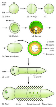
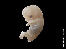
Trong sinh học phát triển, sự phát triển phôi mô tả các giai đoạn của quá trình từ hợp tử đến phôi. Hợp tử là một tế bào lưỡng bội hình thành từ sự dung hợp giữa tế bào trứng đơn bội và tế bào tinh trùng đơn bội.[1] Trong phát triển phôi, hợp tử nguyên phân mà không sinh trưởng tế bào (phân cắt), sau đó biệt hóa để tạo nên phôi đa bào.[2][3] Ở động vật có vú, thuật ngữ này thường chỉ nhắc đến những bước sớm, còn những bước sau đó thuộc sự phát triển bào thai.[2][4]
Những giai đoạn chính của sự phát triển phôi là:
- Phân cắt (cleavage): hợp tử phân chia nhưng không sinh trưởng, tạo thành phôi dâu (morula)
- Sự hình thành phôi nang (blastrulation): Phôi dâu phát triển thành phôi nang (blastrula).
- Sự hình thành phôi vị (gastrulation): Phôi nang phát triển thành phôi vị (gastrula).
- Sự phát sinh cơ quan (organogenesis): Phôi nang tiếp tục phát triển và hình thành các cơ quan.
Sau đó, phôi đi vào giai đoạn phát triển tiếp theo, tùy theo loài mà sẽ phát triển thành bào thai hay ấu trùng.
Sự thụ tinh - hợp tử
[sửa | sửa mã nguồn]The egg cell is generally asymmetric, having an animal pole (future ectoderm). It is covered with protective envelopes, with different layers. The first envelope – the one in contact with the membrane of the egg – is made of glycoproteins and is known as the vitelline membrane (zona pellucida in mammals). Different taxa show different cellular and acellular envelopes englobing the vitelline membrane.[2][5]
Fertilization is the fusion of gametes to produce a new organism. In animals, the process involves a sperm fusing with an ovum, which eventually leads to the development of an embryo. Depending on the animal species, the process can occur within the body of the female in internal fertilization, or outside in the case of external fertilization. The fertilized egg cell is known as the zygote.[2][5]
To prevent more than one sperm fertilizing the egg (polyspermy), fast block and slow block to polyspermy are used. Fast block, the membrane potential rapidly depolarizing and then returning to normal, happens immediately after an egg is fertilized by a single sperm. Slow block begins in the first few seconds after fertilization and is when the release of calcium causes the cortical reaction, in which various enzymes are released from cortical granules in the eggs plasma membrane, causing the expansion and hardening of the outside membrane, preventing more sperm from entering.[6][5]
Giai đoạn phân cắt - phôi dâu
[sửa | sửa mã nguồn]
Phân cắt là giai đoạn hợp tử phân chia nhưng không sinh trưởng, trở thành một nhúm tế bào có kích thước tương đương hợp tử ban đầu. Ít nhất có bốn lần phân chia diễn ra, hình thành phôi dâu là một khối cầu đặc có ít nhất mười sáu tế bào. Trong hình thành phôi ban đầu ở chuột, những tế bào chị em của mỗi lần phân chia vẫn kết nối với nhau trong kì trung gian trông qua cầu liên bào.[7] Những tế bào kế từ giai đoạn này đến giai đoạn phôi vị được gọi là nguyên bào (blastomere). Tùy thuộc vào lượng noãn hoàng trong trứng, sự phân cắt có thể diễn ra hoàn toàn (holoblastic) hoặc không hoàn toàn (meroblastic).[8][9]
Sự phân cắt hoàn toàn diễn ra ở những động vật có ít noãn hoàng,[10] bao gồm người và những động vật có vú khác nhận chất dinh dưỡng từ mẹ (qua nhau thai, hay qua sữa ở thú có túi). Sự phân cắt không hoàn toàn diễn ra ở những động vật có nhiều noãn hoàng, bao gồm chim và bò sát. Vì sự phân cắt bị lệch về phía cực động vật, sự phân bổ về số lượng và kích thước của tế bào không đồng đều trong phôi dâu, thường ở cực động vật có nhiều tế bào và tế bào nhỏ hơn.[8][9]
Trong những trứng phân cắt hoàn toàn, lần phân cắt đầu tiên luôn luôn diễn ra theo trực phân chia cực động vật - cực thực vật, lần phân cắt thứ hai vuông góc với lần phân cắt đầu tiên. Các lần phân cắt sau có thể diễn ra theo nhiều kiểu mẫu, tùy theo động vật.
| Phân cắt hoàn toàn | Phân cắt không hoàn toàn |
|---|---|
|
Cuối giai đoạn phân cắt diễn ra quá trình "chuyển đối giữa phôi nang" xảy ra đồng thời với sự phiên mã của hợp tử.
The end of cleavage is known as midblastula transition and coincides with the onset of zygotic transcription.
In amniotes, the cells of the morula are at first closely aggregated, but soon they become arranged into an outer or peripheral layer, the trophoblast, which does not contribute to the formation of the embryo proper, and an inner cell mass, from which the embryo is developed. Fluid collects between the trophoblast and the greater part of the inner cell-mass, and thus the morula is converted into a vesicle, called the blastodermic vesicle. The inner cell mass remains in contact, however, with the trophoblast at one pole of the ovum; this is named the embryonic pole, since it indicates the location where the future embryo will develop.[18][9]
Giai đoạn phôi nang
[sửa | sửa mã nguồn]Kể từ lần phân cắt thứ bảy (tối đa 128 tế bào), phôi dâu chính thức trở thành phôi nang.[8] Phôi nang gồm bì phôi, bao bọc "khoang phôi" là một khoang chứa dịch hoặc chưa noãn hoàng.[cần dẫn nguồn]
Ở thú, cấu trúc tương đương với phôi nang là túi phôi (blastocyst), lớp ngoài của túi phôi không được gọi là bì phôi mà gọi là "lá nuôi" hay "nguyên bào nuôi" (tropoblast), và bên trong của túi phôi có thêm một khối tế bào gọi là "là phôi" hay "phôi bào" (embroblast).
Trong giai đoạn này, tế bào của nguyên bào nuôi biệt hóa thành hai lớp: lớp hợp bào lá nuôi và lớp tế bào lá nuôi. Lớp hợp bào nuôi là một lớp chất nguyên sinh lấm chấm những nhân, nhưng ngăn cách thành các tế bào (nên vì thế gọi là hợp bào), còn lớp tế bào lá nuôi là các tế bào thật sự. Nguyên bào nuôi không phát triển thành phôi, mà nó phát triển thành màng đệm và đóng vai trò quan trọng trong sự phát triển của nhau thai.
Về phôi bào, trong giai đoạn này nó biệt hóa thành hai lớp: lá phôi trong (hypoblast) và lá phôi ngoài (epiblast). Hai lớp này cấu nên đĩa phôi. Khoảng không ở hai bên đĩa phôi, giới hạn bởi lá phôi được gọi là khoang túi phôi (ở phía lá phôi trong, tức mặt trước) khoang màng ối (ở phía lá phôi ngoài, tức mặt lưng).[18][19] Lá phôi trong là tiền thân của lớp nội bì và lá phôi ngoài là tiền thần của lớp ngoại bì.
Hình thành lớp mầm
[sửa | sửa mã nguồn]
The embryonic disc becomes oval and then pear-shaped, the wider end being directed forward. Towards the narrow, posterior end, an opaque primitive streak, is formed and extends along the middle of the disc for about half of its length; at the anterior end of the streak there is a knob-like thickening termed the primitive node or knot, (known as Hensen's knot in birds). A shallow groove, the primitive groove, appears on the surface of the streak, and the anterior end of this groove communicates by means of an aperture, the blastopore, with the yolk sac. The primitive streak is produced by a thickening of the axial part of the ectoderm, the cells of which multiply, grow downward, and blend with those of the subjacent endoderm. From the sides of the primitive streak a third layer of cells, the mesoderm, extends laterally between the ectoderm and endoderm; the caudal end of the primitive streak forms the cloacal membrane. The blastoderm now consists of three layers, an outer ectoderm, a middle mesoderm, and an inner endoderm; each has distinctive characteristics and gives rise to certain tissues of the body. For many mammals, it is sometime during formation of the germ layers that implantation of the embryo in the uterus of the mother occurs.[18][19]
Giai đoạn phôi vị
[sửa | sửa mã nguồn]Trong giai đoạn phôi vị, tế bào di chuyển đến bên trong phôi nang, hình thành nên hai hoặc ba lớp mầm, tùy vào loài động vật. Phôi trong giai đoạn này được gọi là phôi vị, và ba lớp mầm là ngoại bì (ectoderm), trung bì (mesoderm) và nội bì (endoderm). Động vật hai lớp mầm thì không có lớp trung bì.[8] Ở những động vật khác nhau, tổ hợp khác nhau của những quá trình này sẽ diễn ra ở bên trong phôi vị:
- Mọc phủ (epiboly): một phiến tế bào trải lên trên các tế bào khác[1][9]
- Di nhập (ingression): các tế bào riêng lẻ di chuyển vào trong phôi (tế bào di chuyển bằng chân giả)[2][9]
- Chiết nhập (invagination): phiến tế bào gấp về phía phôi, hình thành miệng, hậu môn và ruột nguyên thủy.[8][9]
- Quyển nhập (involution): lớp tế bào ngoài xoắn vặn của phiến tế bào về phía trong.[9]
- Phân lớp (delamination): một phiến tế bào tách làm hai phiến.[9]
- Một số sự thay đổi lớn khác
- Sự phiên mã kịch liệt của những gene của phôi, trước đó thì chỉ sử dụng RNA của mẹ
- Các tế bào kịch liệt biệt hóa, mất đi tính toàn năng
Ở hầu hết các loài động vật, bào tử chồi đựoc hình thành ở vị trí các tế bào di chuyển vào trong. Dựa vào phái sinh của bào tử chồi, động vật có thể được chia thành hai nhóm: động vật miệng thứ sinh (deuterosome) và động vật miệng nguyên sinh (protosome).[9]
Formation of the early nervous system – neural groove, tube and notochord
[sửa | sửa mã nguồn]In front of the primitive streak, two longitudinal ridges, caused by a folding up of the ectoderm, make their appearance, one on either side of the middle line formed by the streak. These are named the neural folds; they commence some little distance behind the anterior end of the embryonic disk, where they are continuous with each other, and from there gradually extend backward, one on either side of the anterior end of the primitive streak. Between these folds is a shallow median groove, the neural groove. The groove gradually deepens as the neural folds become elevated, and ultimately the folds meet and coalesce in the middle line and convert the groove into a closed tube, the neural tube or canal, the ectodermal wall of which forms the rudiment of the nervous system. After the coalescence of the neural folds over the anterior end of the primitive streak, the blastopore no longer opens on the surface but into the closed canal of the neural tube, and thus a transitory communication, the neurenteric canal, is established between the neural tube and the primitive digestive tube. The coalescence of the neural folds occurs first in the region of the hind brain, and from there extends forward and backward; toward the end of the third week, the front opening (anterior neuropore) of the tube finally closes at the anterior end of the future brain, and forms a recess that is in contact, for a time, with the overlying ectoderm; the hinder part of the neural groove presents for a time a rhomboidal shape, and to this expanded portion the term sinus rhomboidalis has been applied. Before the neural groove is closed, a ridge of ectodermal cells appears along the prominent margin of each neural fold; this is termed the neural crest or ganglion ridge, and from it the spinal and cranial nerve ganglia and the ganglia of the sympathetic nervous system are developed.[cần dẫn nguồn] By the upward growth of the mesoderm, the neural tube is ultimately separated from the overlying ectoderm.[20][9]

The cephalic end of the neural groove exhibits several dilatations that, when the tube is closed, assume the form of the three primary brain vesicles, and correspond, respectively, to the future forebrain (prosencephalon), midbrain (mesencephalon), and hindbrain (rhombencephalon) (Fig. 18). The walls of the vesicles are developed into the nervous tissue and neuroglia of the brain, and their cavities are modified to form its ventricles. The remainder of the tube forms the spinal cord (medulla spinalis); from its ectodermal wall the nervous and neuroglial elements of the spinal cord are developed, while the cavity persists as the central canal.[20][9]
Formation of the early septum
[sửa | sửa mã nguồn]The extension of the mesoderm takes place throughout the whole of the embryonic and extra-embryonic areas of the ovum, except in certain regions. One of these is seen immediately in front of the neural tube. Here the mesoderm extends forward in the form of two crescentic masses, which meet in the middle line so as to enclose behind them an area that is devoid of mesoderm. Over this area, the ectoderm and endoderm come into direct contact with each other and constitute a thin membrane, the buccopharyngeal membrane, which forms a septum between the primitive mouth and pharynx.[18][9]
Early formation of the heart and other primitive structures
[sửa | sửa mã nguồn]In front of the buccopharyngeal area, where the lateral crescents of mesoderm fuse in the middle line, the pericardium is afterward developed, and this region is therefore designated the pericardial area. A second region where the mesoderm is absent, at least for a time, is that immediately in front of the pericardial area. This is termed the proamniotic area, and is the region where the proamnion is developed; in humans, however, it appears that a proamnion is never formed. A third region is at the hind end of the embryo, where the ectoderm and endoderm come into apposition and form the cloacal membrane.[18][9]
Sự hình thành khúc
[sửa | sửa mã nguồn]Somitogenesis is the process by which somites (primitive segments) are produced. These segmented tissue blocks differentiate into skeletal muscle, vertebrae, and dermis of all vertebrates.[21]
Somitogenesis begins with the formation of somitomeres (whorls of concentric mesoderm) marking the future somites in the presomitic mesoderm (unsegmented paraxial). The presomitic mesoderm gives rise to successive pairs of somites, identical in appearance that differentiate into the same cell types but the structures formed by the cells vary depending upon the anteroposterior (e.g., the thoracic vertebrae have ribs, the lumbar vertebrae do not). Somites have unique positional values along this axis and it is thought that these are specified by the Hox homeotic genes.[21]
Toward the end of the second week after fertilization, transverse segmentation of the paraxial mesoderm begins, and it is converted into a series of well-defined, more or less cubical masses, also known as the somites, which occupy the entire length of the trunk on either side of the middle line from the occipital region of the head. Each segment contains a central cavity (known as a [myocoel), which, however, is soon filled with angular and spindle-shape cells. The somites lie immediately under the ectoderm on the lateral aspect of the neural tube and notochord, and are connected to the lateral mesoderm by the intermediate cell mass. Those of the trunk may be arranged in the following groups, viz.: cervical 8, thoracic 12, lumbar 5, sacral 5, and coccygeal from 5 to 8. Those of the occipital region of the head are usually described as being four in number. In mammals, somites of the head can be recognized only in the occipital region, but a study of the lower vertebrates leads to the belief that they are present also in the anterior part of the head and that, altogether, nine segments are represented in the cephalic region.[22][21]
Sự phát sinh cơ quan
[sửa | sửa mã nguồn]
At some point after the different germ layers are defined, organogenesis begins. The first stage in vertebrates is called neurulation, where the neural plate folds forming the neural tube (see above).[8] Other common organs or structures that arise at this time include the heart and somites (also above), but from now on embryogenesis follows no common pattern among the different taxa of the animalia.[2]
In most animals organogenesis, along with morphogenesis, results in a larva. The hatching of the larva, which must then undergo metamorphosis, marks the end of embryonic development.[2]
See also
[sửa | sửa mã nguồn]References
[sửa | sửa mã nguồn]- ^ Gilbert, Scott (2000). Developmental Biology. 6th edition. Chapter 7 Fertilization: Beginning a new organism. Truy cập ngày 3 tháng 10 năm 2020.
- ^ a b c d e f Gilbert, Scott (2000). Developmental Biology. 6th edition. The Circle of Life: The Stages of Animal Development. Truy cập ngày 3 tháng 10 năm 2020.
- ^ Drost, Hajk-Georg; Janitza, Philipp; Grosse, Ivo; Quint, Marcel (2017). “Cross-kingdom comparison of the developmental hourglass”. Current Opinion in Genetics & Development. 45: 69–75. doi:10.1016/j.gde.2017.03.003. PMID 28347942.
- ^ Gilbert, Scott (2000). Developmental Biology. 6th edition. Early Mammalian Development. Truy cập ngày 3 tháng 10 năm 2020.
- ^ a b c Hinton-Sheley, Phoebe. “Stages of Early Embryonic Development”. Truy cập ngày 6 tháng 10 năm 2020.
- ^ Alberts, Bruce; Johnson, Alexander; Lewis, Julian; Raff, Martin; Roberts, Keith; Walter, Peter (2002). “Fertilization” (bằng tiếng Anh). Lưu trữ bản gốc ngày 14 tháng 5 năm 2017.
- ^ Zenker, J.; White, M. D.; Templin, R. M.; Parton, R. G.; Thorn-Seshold, O.; Bissiere, S.; Plachta, N. (tháng 9 năm 2017). “A microtubule-organizing center directing intracellular transport in the early mouse embryo”. Science (bằng tiếng Anh). 357 (6354): 925–928. Bibcode:2017Sci...357..925Z. doi:10.1126/science.aam9335. ISSN 0036-8075. PMID 28860385. S2CID 206658036.
- ^ a b c d e f What is a cell? Lưu trữ 2006-01-18 tại Wayback Machine 2004. A Science Primer: A Basic Introduction to the Science Underlying NCBI Resources. NCBI; and Campbell, Neil A.; Reece, Jane B.; Biology Benjamin Cummings, Pearson Education 2002.
- ^ a b c d e f g h i j k l m n o p q r s t Gilbert, Scott (2000). Developmental Biology. 6th edition. An Introduction of Early Development Process. Truy cập ngày 3 tháng 10 năm 2020.
- ^ Gilbert, Scott (2000). Early Development of the Nematode Caenorhabditis elegans (ấn bản thứ 6). Truy cập ngày 3 tháng 10 năm 2020.
- ^ Gilbert, Scott (2000). Developmental Biology. 6th edition. The Early Development of Sea Urchins. Truy cập ngày 3 tháng 10 năm 2020.
- ^ Gilbert, Scott (2000). Developmental Biology. 6th edition. Early Development in Tunicates. Truy cập ngày 4 tháng 10 năm 2020.
- ^ Gilbert, Scott (2000). Developmental Biology. 6th edition. Early Amphibian Development. Truy cập ngày 3 tháng 10 năm 2020.
- ^ Gilbert, Scott (2000). Developmental Biology. 6th edition. The Early Development of Snails. Truy cập ngày 4 tháng 10 năm 2020.
- ^ a b Gilbert, Scott (2000). Developmental Biology. 6th edition. Chapter 11. The early development of vertebrates: Fish, birds, and mammals. Truy cập ngày 3 tháng 10 năm 2020.
- ^ Gilbert, Scott (2000). Developmental Biology. 6th edition. Early Development of the Nematode Caenorhabditis elegans. Truy cập ngày 4 tháng 10 năm 2020.
- ^ Gilbert, Scott (2000). Developmental Biology. 6th edition. Early Drosophila Development. Truy cập ngày 4 tháng 10 năm 2020.
- ^ a b c d e “Yahoo”. Yahoo. Bản gốc lưu trữ ngày 22 tháng 12 năm 2009.
- ^ a b Balano, Alex (25 tháng 2 năm 2019). “What is the Blastocyst”. Science Trends. Truy cập ngày 5 tháng 10 năm 2020.
- ^ a b “The Neural Groove and Tube”. Yahoo. Bản gốc lưu trữ ngày 22 tháng 8 năm 2007.
- ^ a b c Pourquié, Oliver (tháng 11 năm 2001). “Vertebrate Somitogenesis”. Annual Review of Cell and Developmental Biology. 17: 311–350. doi:10.1146/annurev.cellbio.17.1.311. PMID 11687492. Truy cập ngày 5 tháng 10 năm 2020.
- ^ “The Primitive Segments”. Yahoo. Bản gốc lưu trữ ngày 11 tháng 9 năm 2007.
External links
[sửa | sửa mã nguồn]- Cellular Darwinism
- Embryogenesis & MMPs, PMAP The Proteolysis Map-animation
- Development of the embryo (retrieved November 20, 2007)
- Video of embryogenesis of the frog Xenopus laevis from shortly after fertilization until the hatching of the tadpole; acquired by MRI (DOI of paper)
