Thành viên:Naazulene/Ngoại bì
| Ectoderm | |
|---|---|
 Organs derived from ectoderm. | |
 Section through embryonic disk of Vespertilio murinus. | |
| Chi tiết | |
| Các ngày | 16 |
| Thuật ngữ giải phẫu | |
Ngoại bì (tiếng Anh: ectoderm) là lớp ngoài cùng trong ba lớp mầm của phôi nguyên thủy, dưới nó là trung bì (lớp giữa) và nội bì (lớp trong cùng).[1] Nó bắt nguồn từ lớp ngoài cùng của tế bào mầm.[2]
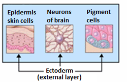
Nhìn chung, ngoại bì sẽ phân hóa thành mô thần kinh (não, tủy sống và dây thần kinh) và biểu mô (da, miệng, hậu hôn, lỗ mũi, da, móng,[3] men răng). Một số loại biểu mô được phân hóa từ nội bì.[3]
Trong mô của sinh vật có xương sống, ngoại bì được chia thành hai phần: bề lưng ngoài bì - còn được gọi là ngoại bì ngoài, và tấm thần kinh (neural plate). Tấm thần kinh lõm vào tạo thành ống thần kinh và mào thần kinh.[4] Vì vậy, tấm thần kinh và mào thần kinh còn được gọi là ngoại bì thần kinh.
Lịch sử
[sửa | sửa mã nguồn]Heinz Christian Pander, một nhà sinh học người Đức Baltic được ghi nhận cho phát hiện về ba lớp mầm của phôi nguyên thủy. Pander nhận bằng tiến sĩ động vật học từ Đại học Würzburg vào năm 1817. Ông bắt đầu nghiên cứu về phôi học sử dụng trứng gà, từ đó ông phát hiện ra ngoại bì, trung bì, và nội bì. Vì những phát hiện này, ông đôi khi được gọi là "cha đẻ của phôi học".
Công trình nghiên cứu phôi nguyên thủy của Pander được tiếp nối bởi một nhà sinh học người Prussian–Estonian tên Karl Ernst von Baer. Qua nhiều nghiên cứu lên nhiều loài khác nhau, Baer đã mở rộng ý tưởng của Pander lên tất cả động vật có xương sống. Baer cũng được ghi nhận cho phát hiện túi phôi (blastula). Năm 1828, những nghiên cứu của Baer được ông xuất bản trong cuốn sách "On the Development of Animal" (tạm dịch: Về sự Phát triển của Động vật).[5]
Biệt hóa
[sửa | sửa mã nguồn]Hình mạo ban đầu
[sửa | sửa mã nguồn]Lớp ngoại bị có thể được quan sát ở lưỡng cư và cá trong những pha sau của giai đoạn phôi vị (gastrulation). Giai đoạn phôi vị bắt đầu khi phôi phân chia thành nhiều tế bào, tạo thành khối hình cầu rỗng gọi là phôi nang. Phôi nang lúc này phân cực, có hai cực là cực động vật và cực thực vật. Tiền thần của ngoại bì nằm ở cực động vật.[2]
Sự phát triển ban đầu
[sửa | sửa mã nguồn]Giống như hai lớp mầm còn lại - lớp trung bì và lớp nội bì - lớp ngoại bì hình thành nhanh chóng sau thời điểm thụ tinh. Vị trí tương đối của lớp ngoại bì trong phôi được quyết định bởi "ái lực chọn lọc" của nó: bề trong của lớp ngoại bì có ái lực mạnh/dương đối với lớp trung bì, và ái lực yếu/âm đối với lớp nội bì.[6] Ái lực chọn lọc này thay đổi trong từng giai đoạn của phát triển phôi. Cường độ của ái lực giữa hai bề mặt của hai lớp mầm được quyết định bởi lượng là loại phân tử cadherin ở bề mặt tế bào. Ví dụ, sự biểu hiện của N-cadherin quan trọng để đảm bảo sự tách biệt của tiền thân tế bào thần kinh với tiền thân tế bào biểu mô.[2] Tương tự, trong khi bề
Like the other two germ layers – i.e., the mesoderm and endoderm – the ectoderm forms shortly after fertilization, after which rapid cell division begins. The position of the ectoderm relative to the other germ layers of the embryo is governed by "selective affinity", meaning that the inner surface of the ectoderm has a strong (positive) affinity for the mesoderm, and a weak (negative) affinity for the endoderm layer.[6] This selective affinity changes during different stages of development. The strength of the attraction between two surfaces of two germ layers is determined by the amount and type of cadherin molecules present on the cells' surface. For example, the expression of N-cadherin is crucial to maintaining separation of precursor neural cells from precursor epithelial cells.[2] Likewise, while the surface ectoderm becomes the epidermis,[6] the neuroectoderm is induced along the neural pathway by the notochord, which is typically positioned above it.[2][4]
Giai đoạn phôi vị
[sửa | sửa mã nguồn]Trong giai đoạn phôi vị, tế bào hình chai chiết nhập vào mặt lưng của của phôi nang để hình thành miệng phôi (blastopore). Các tế bào tiếp tục lõm vào, hình thành khoang phôi (blastocoel) là khoảng không giữa vách phôi nang và vách của miệng phôi mới hình thành.
During the process of gastrulation, bottle cells invaginate on the dorsal surface of the blastula to form the blastopore. The cells continue to extend inward and migrate along the inner wall of the blastula to form a fluid-filled cavity called the blastocoel. The once superficial cells of the animal pole are destined to become the cells of the middle germ layer called the mesoderm. Through the process of radial extension, cells of the animal pole that were once several layers thick divide to form a thin layer. At the same time, when this thin layer of dividing cells reaches the dorsal lip of the blastopore, another process occurs termed convergent extension. During convergent extension, cells that approach the lip intercalate mediolaterally, in such a way that cells are pulled over the lip and inside the embryo. These two processes allow for the prospective mesoderm cells to be placed between the ectoderm and the endoderm. Once convergent extension and radial intercalation are underway, the rest of the vegetal pole, which will become endoderm cells, is completely engulfed by the prospective ectoderm, as these top cells undergo epiboly, where the ectoderm cells divide in a way to form one layer. This creates a uniform embryo composed of the three germ layers in their respective positions.[2]
Sự phát triển sau
[sửa | sửa mã nguồn]Sau khi ba lớp mầm đã được hình thành, sự biệt hóa tế bào chính thức diễn ra.
Một trong những quá trình quan trọng ở đây là sự hình thành ống thần kinh (neurulation), trong đó lớp ngoại bì biệt hóa thành ống thần kinh, mào thần kinh và biểu bì. Mỗi thành phần này sẽ phát triển lên thành một nhóm các cơ quan: ống thần kinh phát triển thành hệ thần kinh trung ương; mào thần kinh phát triển thành hệ thần kinh ngoại biên, tế bào hắc tố (melanocyte) và sụn mặt; biểu bì phát triển thành biểu mô, lông tóc, móng, tuyến bã, \
Once the three germ layers have been established, cellular differentiation can occur. The first major process here is neurulation, wherein the ectoderm differentiates to form the neural tube, neural crest cells and the epidermis. Each of these three components will give rise to a particular complement of cells. The neural tube cells give rise to the central nervous system, neural crest cells give rise to the peripheral and enteric nervous system, melanocytes, and facial cartilage, and the epidermal region will give rise to the epidermis, hair, nails, sebaceous glands, olfactory and oral epithelium, and eyes.[2]
Neurulation
[sửa | sửa mã nguồn]Neurulation occurs in two parts, primary and secondary neurulation. Both processes position neural crest cells between a superficial epidermal layer and the deep neural tube. During primary neurulation, the notochord cells of the mesoderm signal the adjacent, superficial ectoderm cells to reposition themselves into a columnar pattern to form cells of the ectodermal neural plate.[7] As the cells continue to elongate, a group of cells immediately above the notochord change their shape, forming a wedge in the ectodermal region. These special cells are called medial hinge cells (MHPs). As the ectoderm continues to elongate, the ectodermal cells of the neural plate fold inward. The inward folding of the ectoderm by virtue of mainly cell division continues until another group of cells forms within the neural plate. These cells are termed dorsolateral hinge cells (DLHPs), and, once formed, the inward folding of the ectoderm stops. The DLHP cells function in a similar fashion as MHP cells regarding their wedge like shape, however, the DLHP cells result in the ectoderm converging. This convergence is led by ectodermal cells above the DLHP cells known as the neural crest. The neural crest cells eventually pull the adjacent ectodermal cells together, which leaves neural crest cells between the prospective epidermis and hollow, neural tube.[2]
Organogenesis
[sửa | sửa mã nguồn]
All of the organs that rise from the ectoderm such as the nervous system, teeth, hair and many exocrine glands, originate from two adjacent tissue layers: the epithelium and the mesenchyme.[8] Several signals mediate the organogenesis of the ectoderm such as: FGF, TGFβ, Wnt, and regulators from the hedgehog family. The specific timing and manner that the ectodermal organs form is dependent on the invagination of the epithelial cells.[9] FGF-9 is an important factor during the initiation of tooth germ development. The rate of epithelial invagination in significantly increased by action of FGF-9, which is only expressed in the epithelium, and not in the mesenchyme. FGF-10 helps to stimulate epithelial cell proliferation, in order make larger tooth germs. Mammalian teeth develop from ectoderm derived from the mesenchyme: oral ectoderm and neural crest. The epithelial components of the stem cells for continuously growing teeth form from tissue layers called the stellate reticulum and the suprabasal layer of the surface ectoderm.[9]
Ý nghĩa lâm sàng
[sửa | sửa mã nguồn]Loạn sản ngoại bì
[sửa | sửa mã nguồn]Loạn sản ngoại bì (ectodermal dysplasia) là bệnh nghiêm trọng trong đó những tế bào có nguồn gốc ngoại bì phát triển một cách bất thường. Có hơn 170 loại loạn sản ngoại bì. Bệnh này gây ra bởi một đột biến hay một tổ hợp đột biến trong một số gen nhất định. Nghiên cứu về loạn sản ngoại bì vẫn đang được thực hiện, vì người ta mới chỉ nhận diện được một số nhỏ những đột biến liên quan đến một loại loạn sản ngoại bì.[10]
Loạn sản ngoại bì thiếu tuyến mồ hôi (HED) là loại phổ biến nhất. Triệu chứng đặc trưng nhất của bệnh này là cơ thể không sản xuất đủ mồ hôi do không có tuyến mồ hôi hoặc tuyến mồ hôi bị hư hỏng. Vì đổ mồ hôi là cơ chế hạ nhiệt của cơ thể, người mắc bệnh này sẽ bị giới hạn khả năng chơi thể thao, gặp khó khăn với mùa hè. Người mắc bệnh này ở vùng khí hậu ấm nóng có thể tử vong vì sốc nhiệt. HED cũng cơ thể gây ra dị dạng ở mặt, ví dụ như răng nhọn hoặc không có răng, da nhăn nheo ở vùng quanh mắt, mũi dị hình, sẹo, tóc mỏng. Vấn đề về da như chàm da cũng đã được quan sát ở một số ca. Hầu hết bệnh nhân mang đột biếngen EDA trên nhiễm sắc thể X.[11] Vì alen gây bệnh thường là alen lặn trên nhiễm sắc thể X, bệnh này xuất hiện ở nam nhiều hơn ở nữ.

Xem thêm
[sửa | sửa mã nguồn]- Sự biệt hóa của ngoại bì
- Coelom
- Phôi học
- Ngoại bì
- Giai đoạn phôi nang
References
[sửa | sửa mã nguồn]- ^ Langman's Medical Embryology, 11th edition. 2010.
- ^ a b c d e f g h Gilbert, Scott F. Developmental Biology. 9th ed. Sunderland, MA: Sinauer Associates, 2010: 333-370. Print.
- ^ a b “Derivation of Tissues | SEER Training”. training.seer.cancer.gov.
- ^ a b Marieb, Elaine N.; Hoehn, Katja (2019). Human Anatomy & Physiology. United States of America: Pearson. tr. 146, 482–483, 1102–1106. ISBN 978-0-13-458099-9.
- ^ Baer KE von (1986) In: Oppenheimer J (ed.) and Schneider H (transl.), Autobiography of Dr. Karl Ernst von Baer. Canton, MA: Science History Publications.
- ^ a b c Hosseini, Hadi S.; Garcia, Kara E.; Taber, Larry A. (2017). “A new hypothesis for foregut and heart tube formation based on differential growth and actomyosin contraction”. Development. 144 (13): 2381–2391. doi:10.1242/dev.145193. PMC 5536863. PMID 28526751.
- ^ O'Rahilly, R; Müller, F (1994). “Neurulation in the Normal Human Embryo”. Ciba Foundation Symposium 181 - Neural Tube Defects. Ciba Foundation Symposium. Novartis Foundation Symposia. 181. tr. 70–82. doi:10.1002/9780470514559.ch5. ISBN 9780470514559. PMID 8005032.
- ^ Pispa, J; Thesleff, I (15 tháng 10 năm 2003). “Mechanisms of ectodermal organogenesis”. Developmental Biology. 262 (2): 195–205. doi:10.1016/S0012-1606(03)00325-7. PMID 14550785.
- ^ a b Tai, Y. Y.; Chen, R. S.; Lin, Y.; Ling, T. Y.; Chen, M. H. (2012). “FGF-9 accelerates epithelial invagination for ectodermal organogenesis in real time bioengineered organ manipulation”. Cell Communication and Signaling. 10 (1): 34. doi:10.1186/1478-811X-10-34. PMC 3515343. PMID 23176204.
- ^ Priolo, M.; Laganà, C (tháng 9 năm 2001). “Ectodermal Dysplasias: A New Clinical-Genetic Classification”. Journal of Medical Genetics. 38 (9): 579–585. doi:10.1136/jmg.38.9.579. PMC 1734928. PMID 11546825.
- ^ Bayes, M.; Hartung, A. J.; Ezer, S.; Pispa, J.; Thesleff, I.; Srivastava, A. K.; Kere, J. (1998). “The Anhidrotic Ectodermal Dysplasia Gene (EDA) Undergoes Alternative Splicing and Encodes Ectodysplasin-A with Deletion Mutations in Collagenous Repeats”. Human Molecular Genetics. 7 (11): 1661–1669. doi:10.1093/hmg/7.11.1661. PMID 9736768.
