Thành viên:Naazulene/Chi trên
| Upper limb | |
|---|---|
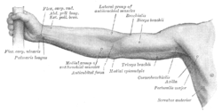
 Front of right upper extremity. | |
 Back of right upper extremity. | |
| Chi tiết | |
| Cơ quan | Musculoskeletal |
| Định danh | |
| Latinh | membrum superius |
| Thuật ngữ giải phẫu | |
Chi trên là tay của động vật bốn chân đứng thẳng, kéo dài từ xương vai, xương ức đến các xương đốt ngón tay, bao gồm các cơ và dây chằng trong vai, khuỷu, cổ tay và các đốt ngón tay.[1] Ở người, vùng chi trên được chia thành vai, cánh tay, cẳng tay, cổ tay, và bàn tay.[2] Công dụng chính của nó là leo trèo, nâng và cầm nắm vật dụng. Trong giải phẫu, chữ cánh tay chỉ bao gồm phần giữa vai và khuỷu, còn trong ngôn ngữ bình thường thì cánh tay là phần giữa vai và cổ tay, hoặc thậm chí là đến bàn tay.
Cấu trúc
[sửa | sửa mã nguồn]Trong cơ thể người, các cơ của vùng chi trên có thể được phân loại dựa nguyên ủy, hình dáng, chức năng hay thần kinh chi phối. Cách phân loại theo thần kinh chi phối giúp nghiên cứu nguồn gốc phôi học là tiến hóa, còn cách phân loại chức năng - hình dáng thể hiện các điểm giống nhau giữa cơ chế hoạt động của các cơ.[3]
Xương
[sửa | sửa mã nguồn]Các xương của mỗi vùng chi trên bao gồm:
| Xương | Số lượng (mỗi bên) | Phân loại | |
|---|---|---|---|
| Đai vai | Xương đòn | 1 | Xương dài |
| Xương vai | 1 | Xương dẹt | |
| Cánh tay | Xương cánh tay | 1 | Xương dài |
| Cẳng tay | Xương quay | 1 | Xương dài |
| Xương trụ | 1 | Xương dài | |
| Cổ tay | Xương thuyền | 1 | Xương khác |
| Xương nguyệt | 1 | Xương khác | |
| Xương tháp | 1 | Xương khác | |
| Xương đậu | 1 | Xương khác | |
| Xương thang | 1 | Xương khác | |
| Xương thê | 1 | Xương khác | |
| Xương cả | 1 | Xương khác | |
| Xương móc | 1 | Xương khác | |
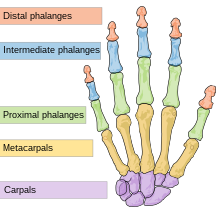
| Bàn tay | Xương đốt bàn | 5 | Xương dài |
| Xương đốt ngón | 14 | Xương dài |
Các khớp của xương chi trên bao gồm
| Tên khớp | Tên xương | |
|---|---|---|
| Khớp cùng đòn | Xương vai | Xương đòn |
| Khớp ức đòn | Xương ức | Xương đòn |
| Khớp vai | Xương vai | Xương cánh tay |
| Khớp khuỷu | Xương cánh tay | Xương quay và xương trụ |
| Khớp quay trụ trên | Xương quay | Xương trụ |
| Khớp quay trụ dưới | Xương quay | Xương trụ |
| Khớp quay cổ tay | Xương quay | Các xương cổ tay |
| Các khớp nhỏ | Xương cổ tay | Xương đốt bàn |
| Xương đốt bàn | Xương đốt ngón | |
| Xương đốt ngón | Xương đốt ngón | |
Đai vai
[sửa | sửa mã nguồn]
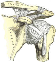
Đai vai[4][5] bao gồm xương ức và xương vai, nó nối chi trên vào hệ xương trục ở khớp ức đòn. Khớp này là một bao hoạt dịch được trợ lực bởi cơ dưới đòn, cơ dưới đòn hoạt động như một dây chằng động. Cơ này giúp tránh trật khớp, và những tác động mạch lên đây sẽ làm gãy xương đòn. Khớp cùng đòn - khớp giữa mỏm cùng vai trên xương vai và diện cùng vai trên xương đòn - cũng được trợ lực bởi nhiều dây chằng mạnh, đặc biệt là dây chằng quạ đòn giúp hạt chế cử động quá mức. Hai khớp này cho phép đai vai thực hiện rất nhiều nhiều cử động. Mặt khác, đai hông không thể thực hiện nhiều cử động nhưng lại có tính ổn định và khả năng chịu lực cao vì nó được gắn chặt vào hệ xương trục.[5]
Cử động của đai vai được trợ lực bởi nhiều cơ. Cơ quan trọng nhất là những cơ dạng phiến, thay vì cơ dạng thoi. Các cơ này không bao giờ hoạt động riêng lẻ mà luôn kéo cơ khác hoạt động cùng.[5] Các cơ của vai thường được nahwcs đến là cơ của thân mình.
Các cơ của vai, chưa tính khớp vai, là:
| Tên cơ | Nguyên ủy | Bám tận | |
|---|---|---|---|
| Từ vùng đầu | Cơ thang | xương chẩm | gai vai, các đốt sống |
| Cơ ức đòn chũm | mõm chũm của xương thái dương | xương đòn, xương ức | |
| Cơ vai móng | xương móng | xương đòn | |
| Sau | Cơ trám lớn | các đốt sống T2 - T5 | cạnh trong xương vai, dưới gai |
| Cơ trám bé | các đốt sống C7 - T11 | cạnh trong xương vai, ở gai | |
| Cơ nâng vai | cát đốt sống C1 - C4 | cạnh trong xương vai, trên gai | |
| Trước | Cơ dưới đòn | xương sườn thứ nhất | rãnh dưới đòn của xương đòn |
| Cơ ngực bé | xương sườn thứ ba, bốn và năm | mỏm quạ của xương vai | |
| Cơ răng trước | 8 hoặc 9 xương sườn đầu tiên | cạnh ngoài xương vai |
Khớp vai
[sửa | sửa mã nguồn]
Khớp vai là khớp ổ và bi giữa ổ chảo của xương vai và chỏm của xương cánh tay. Khớp này không được trợ lực thụ động như ở những khớp khác, mà được trợ lực chủ động bởi chóp xoay - nhóm bốn cơ ngắn nối giữa xương vai và xương cánh tay.[6] Khớp vai dễ bị trật về phía trước nhất, chiếm đến khoảng 97%.[7]
Các cơ của khớp vai là:
| Tên cơ | Nguyên ủy | Bám tận | |
|---|---|---|---|
| Mặt sau | Cơ trên gai | Hố trên gai của xương vai | Củ lớn của xương cánh tay |
| Cơ dưới gai | Hố dưới gai của xương vai | Củ lớn của xương cánh tay | |
| Cơ tròn bé | Cạnh ngoài của xương vai | Củ lớn của xương cánh tay | |
| Cơ dưới vai | Góc dưới của xương vai | Bề trong của xương cánh tay | |
| Cơ delta | Gai vai của xương vai, cạnh trước của xương đòn | Lồi củ delta của xương cánh tay | |
| Cơ lưng rộng | Xương chẩm, các đốt sống | Gai vai của xương vai, cạnh trong của xương đòn | |
| Cơ tròn lớn | Cạnh dưới xương vai | Bề trong của xương cánh tay |
Các cơ ở vai hoạt động khá phức tạp, những cử động tượng chừng đơn giản lại cần các cơ khác nhau thực hiện các cử động tương hỗ. Ví dụ, cơ ngực lớn là để gấp vai (đưa tay về trước), cơ lưng rộng là để duỗi vai (đưa tay về sau); khi hoạt động cùng nhau, nó làm động tác xoay vai ra ngoài (như khi mở cửa có bản lề).[6]

Cánh tay
[sửa | sửa mã nguồn]
Cánh tay trong giải phẫu được tính là vùng từ vai đến khuỷu tay. Nó bao gồm xương cánh tay và khớp khuỷu.
Khớp khuỷu bao gồm ba khớp: khớp cánh tay quay, khớp cánh tay trụ và khớp quay trụ trên. Hai khớp đầu tiên tham gia cử động gấp và duỗi khuỷu tay, còn khớp thứ ba tham gia cử động sấp và ngửa cẳng tay, cùng với khớp quay trụ dưới. Cơ gấp chính là cơ nhị đầu cánh tay và cơ duỗi chính là cơ ba đầu cánh tay. Cơ nhị dầu cánh tay cũng là cơ sấp chính.[8]
Các cơ của cánh tay bao gồm:[3]
| Tên cơ | Nguyên ủy | Bám tận | |
|---|---|---|---|
| Mặt sau | Cơ tam đầu cánh tay | Củ dưới ổ chảo của xương vai (đầu dài), trên rãnh quay (đầu ngoài), dưới rãnh quay (đầu trong) | Mỏm cùng vai của xương trụ |
| Cơ khuỷu | Mỏm trên lồi cầu ngoài của xương cánh tay | Mỏm cùng vai của xương trụ | |
| Mặt trước | Cơ cánh tay | ||
| Cơ nhị đầu cánh tay |
Cẳng tay
[sửa | sửa mã nguồn]
The forearm (
tiếng Latinh: antebrachium),[4] composed of the radius and ulna; the latter is the main distal part of the elbow joint, while the former composes the main proximal part of the wrist joint.
Most of the large number of muscles in the forearm are divided into the wrist, hand, and finger extensors on the dorsal side (back of hand) and the ditto flexors in the superficial layers on the ventral side (side of palm). These muscles are attached to either the lateral or medial epicondyle of the humerus. They thus act on the elbow, but, because their origins are located close to the centre of rotation of the elbow, they mainly act distally at the wrist and hand. Exceptions to this simple division are brachioradialis — a strong elbow flexor — and palmaris longus — a weak wrist flexor which mainly acts to tense the palmar aponeurosis. The deeper flexor muscles are extrinsic hand muscles; strong flexors at the finger joints used to produce the important power grip of the hand, whilst forced extension is less useful and the corresponding extensor thus are much weaker. [9]
Biceps is the major supinator (drive a screw in with the right arm) and pronator teres and pronator quadratus the major pronators (unscrewing) — the latter two role the radius around the ulna (hence the name of the first bone) and the former reverses this action assisted by supinator. Because biceps is much stronger than its opponents, supination is a stronger action than pronation (hence the direction of screws). [9]
- Muscles
- of the forearm[3]
- Posterior
- (Superficial) extensor digitorum, extensor digiti minimi, extensor carpi ulnaris, (deep) supinator, abductor pollicis longus, extensor pollicis brevis, extensor pollicis longus, extensor indicis
- Anterior
- (Superficial) pronator teres, flexor digitorum superficialis, flexor carpi radialis, flexor carpi ulnaris, palmaris longus, (deep) flexor digitorum profundus, flexor pollicis longus, pronator quadratus
- Radial
- Brachioradialis, extensor carpi radialis longus, extensor carpi radialis brevis
Wrist
[sửa | sửa mã nguồn]The wrist (
tiếng Latinh: carpus),[4] composed of the carpal bones, articulates at the wrist joint (or radiocarpal joint) proximally and the carpometacarpal joint distally. The wrist can be divided into two components separated by the midcarpal joints. The small movements of the eight carpal bones during composite movements at the wrist are complex to describe, but flexion mainly occurs in the midcarpal joint whilst extension mainly occurs in the radiocarpal joint; the latter joint also providing most of adduction and abduction at the wrist. [10]

How muscles act on the wrist is complex to describe. The five muscles acting on the wrist directly — flexor carpi radialis, flexor carpi ulnaris, extensor carpi radialis, extensor carpi ulnaris, and palmaris longus — are accompanied by the tendons of the extrinsic hand muscles (i.e. the muscles acting on the fingers). Thus, every movement at the wrist is the work of a group of muscles; because the four primary wrist muscles (FCR, FCU, ECR, and ECU) are attached to the four corners of the wrist, they also produce a secondary movement (i.e. ulnar or radial deviation). To produce pure flexion or extension at the wrist, these muscle therefore must act in pairs to cancel out each other's secondary action. On the other hand, finger movements without the corresponding wrist movements require the wrist muscles to cancel out the contribution from the extrinsic hand muscles at the wrist. [10]
Hand
[sửa | sửa mã nguồn]
The hand (
tiếng Latinh: manus),[4] the metacarpals (in the hand proper) and the phalanges of the fingers, form the metacarpophalangeal joints (MCP, including the knuckles) and interphalangeal joints (IP).
Of the joints between the carpus and metacarpus, the carpometacarpal joints, only the saddle-shaped joint of the thumb offers a high degree of mobility while the opposite is true for the metacarpophalangeal joints. The joints of the fingers are simple hinge joints. [10]
The primary role of the hand itself is grasping and manipulation; tasks for which the hand has been adapted to two main grips — power grip and precision grip. In a power grip an object is held against the palm and in a precision grip an object is held with the fingers, both grips are performed by intrinsic and extrinsic hand muscles together. Most importantly, the relatively strong thenar muscles of the thumb and the thumb's flexible first joint allow the special opposition movement that brings the distal thumb pad in direct contact with the distal pads of the other four digits. Opposition is a complex combination of thumb flexion and abduction that also requires the thumb to be rotated 90° about its own axis. Without this complex movement, humans would not be able to perform a precision grip. [11]
In addition, the central group of intrinsic hand muscles give important contributions to human dexterity. The palmar and dorsal interossei adduct and abduct at the MCP joints and are important in pinching. The lumbricals, attached to the tendons of the flexor digitorum profundus (FDP) and extensor digitorum communis (FDC), flex the MCP joints while extending the IP joints and allow a smooth transfer of forces between these two muscles while extending and flexing the fingers. [11]
- Muscles
- of the hand[3]
Neurovascular system
[sửa | sửa mã nguồn]Nerve supply
[sửa | sửa mã nguồn]
The motor and sensory supply of the upper limb is provided by the brachial plexus which is formed by the ventral rami of spinal nerves C5-T1. In the posterior triangle of the neck these rami form three trunks from which fibers enter the axilla region (armpit) to innervate the muscles of the anterior and posterior compartments of the limb. In the axilla, cords are formed to split into branches, including the five terminal branches listed below. [12] The muscles of the upper limb are innervated segmentally proximal to distal so that the proximal muscles are innervated by higher segments (C5–C6) and the distal muscles are innervated by lower segments (C8–T1). [13]
Motor innervation of upper limb by the five terminal nerves of the brachial plexus:[13]
- The musculocutaneous nerve innervates all the muscles of the anterior compartment of the arm.
- The median nerve innervates all the muscles of the anterior compartment of the forearm except flexor carpi ulnaris and the ulnar part of the flexor digitorum profundus. It also innervates the three thenar muscles and the first and second lumbricals.
- The ulnar nerve innervates the muscles of the forearm and hand not innervated by the median nerve.
- The axillary nerve innervates the deltoid and teres minor.
- The radial nerve innervates the posterior muscles of the arm and forearm
Collateral branches of the brachial plexus:[13]
- The dorsal scapular nerve innervates rhomboid major, minor and levator scapulae .
- The long thoracic nerve innervates serratus anterior.
- The suprascapular nerve innervates supraspinatus and infraspinatus
- The lateral pectoral nerve innervates pectoralis major
- The medial pectoral nerve innervates pectoralis major and minor
- The upper subscapular nerve innervates subscapularis
- The thoracodorsal nerve innervates latissimus dorsi
- The lower subscapular nerve innervates subscapularis and teres major
- The medial brachial cutaneous nerve innervates the skin of medial arm
- The medial antebrachial cutaneous nerve innervates the skin of medial forearm
Blood supply and drainage
[sửa | sửa mã nguồn]Arteries of the upper limb:
- The superior thoracic, thoracoacromial, posterior circumflex humeral and subscapular branches of the axillary artery.
- The deep brachial, superior ulnar collateral, inferior ulnar collateral, radial,
ulnar, nutrient and muscular branches of the brachial artery.
- The radial recurrent, muscular, superficial palmar, dorsal carpal, princeps pollicis and radialis indicis branches of the radial artery.
- The anterior ulnar recurrent, posterior ulnar recurrent, anterior interosseous, posterior interosseous and superficial branches of the ulnar artery.

Veins of the upper limb:
As for the upper limb blood supply, there are many anatomical variations.[14]
Other animals
[sửa | sửa mã nguồn]Evolutionary variation
[sửa | sửa mã nguồn]Nội dung của section hầu như chỉ dựa vào một nguồn duy nhất. (July 2011) |

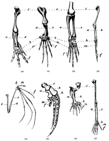
The skeletons of all mammals are based on a common pentadactyl ("five-fingered") template but optimised for different functions. While many mammals can perform other tasks using their forelimbs, their primary use in most terrestrial mammals is one of three main modes of locomotion: unguligrade (hoof walkers), digitigrade (toe walkers), and plantigrade (sole walkers). Generally, the forelimbs are optimised for speed and stamina, but in some mammals some of the locomotion optimisation have been sacrificed for other functions, such as digging and grasping. [15]
In primates, the upper limbs provide a wide range of movement which increases manual dexterity. The limbs of chimpanzees, compared to those of humans, reveal their different lifestyle. The chimpanzee primarily uses two modes of locomotion: knuckle-walking, a style of quadrupedalism in which the body weight is supported on the knuckles (or more properly on the middle phalanges of the fingers), and brachiation (swinging from branch to branch), a style of bipedalism in which flexed fingers are used to grasp branches above the head. To meet the requirements of these styles of locomotion, the chimpanzee's finger phalanges are longer and have more robust insertion areas for the flexor tendons while the metacarpals have transverse ridges to limit dorsiflexion (stretching the fingers towards the back of the hand). The thumb is small enough to facilitate brachiation while maintaining some of the dexterity offered by an opposable thumb. In contrast, virtually all locomotion functionality has been lost in humans while predominant brachiators, such as the gibbons, have very reduced thumbs and inflexible wrists. [15]
In ungulates the forelimbs are optimised to maximize speed and stamina to the extent that the limbs serve almost no other purpose. In contrast to the skeleton of human limbs, the proximal bones of ungulates are short and the distal bones long to provide length of stride; proximally, large and short muscles provide rapidity of step. The odd-toed ungulates, such as the horse, use a single third toe for weight-bearing and have significantly reduced metacarpals. Even-toed ungulates, such as the giraffe, uses both their third and fourth toes but a single completely fused phalanx bone for weight-bearing. Ungulates whose habitat does not require fast running on hard terrain, for example the hippopotamus, have maintained four digits. [15]
In species in the order Carnivora, some of which are insectivores rather than carnivores, the cats are some of the most highly evolved predators designed for speed, power, and acceleration rather than stamina. Compared to ungulates, their limbs are shorter, more muscular in the distal segments, and maintain five metacarpals and digit bones; providing a greater range of movements, a more varied function and agility (e.g. climbing, swatting, and grooming). Some insectivorous species in this order have paws specialised for specific functions. The sloth bear uses their digits and large claws to tear logs open rather than kill prey. Other insectivorous species, such as the giant and red pandas, have developed large sesamoid bones in their paws that serve as an extra "thumb" while others, such as the meerkat, uses their limbs primary for digging and have vestigial first digits. [15]
The arboreal two-toed sloth, a South American mammal in the order Pilosa, have limbs so highly adapted to hanging in branches that it is unable to walk on the ground where it has to drag its own body using the large curved claws on its foredigits. [15]
See also
[sửa | sửa mã nguồn]Notes
[sửa | sửa mã nguồn]- ^ “Upper Extremity”. MeSH. Truy cập ngày 26 tháng 6 năm 2011.
- ^ https://www.kenhub.com/en/library/anatomy/upper-extremity-anatomy
- ^ a b c d Ross & Lamperti 2006, tr. 256
- ^ a b c d Ross & Lamperti 2006, tr. 208
- ^ a b c Sellers 2002, tr. 1–3
- ^ a b Sellers 2002, tr. 3–5
- ^ Abrams, Rachel; Akbarnia, Halleh (2024), “Shoulder Dislocations Overview”, StatPearls, Treasure Island (FL): StatPearls Publishing, PMID 29083735, truy cập ngày 13 tháng 10 năm 2024
- ^ Sellers 2002, tr. 5
- ^ a b Sellers 2002, tr. 6–7
- ^ a b c Sellers 2002, tr. 8–9
- ^ a b Sellers 2002, tr. 10–11
- ^ Seiden 2002, tr. 243
- ^ a b c Seiden 2002, tr. 233–36
- ^ Konarik M, Musil V, Baca V, Kachlik D. Upper limb principal arteries variations: A cadaveric study with terminological implication. Bosn J of Basic Med Sci. 2020;20(4):502-13. DOI: https://doi.org/10.17305/bjbms.2020.4643 PMID 32343941 PMCID: PMC7664784
- ^ a b c d e Gough-Palmer, Maclachlan & Routh 2008, tr. 502–510
References
[sửa | sửa mã nguồn]- Gough-Palmer, Antony L; Maclachlan, Jody; Routh, Andrew (tháng 3 năm 2008). “Paws for Thought: Comparative Radiologic Anatomy of the Mammalian Forelimb” (PDF). RadioGraphics. 28 (2): 501–510. doi:10.1148/rg.282075061. PMID 18349453.
- Ross, Lawrence M; Lamperti, Edward D biên tập (2006). Thieme Atlas of Anatomy: General Anatomy and Musculoskeletal System. Thieme. ISBN 1-58890-419-9.
- Sellers, Bill (2002). “Functional Anatomy of the Upper Limb”. Truy cập ngày 19 tháng 6 năm 2011.
- Seiden, David (2002). USMLE Step 1 Anatomy Notes. Kaplan Medical.
Bản mẫu:Human anatomical features Bản mẫu:Bones of upper extremity Bản mẫu:Joints of upper limbs Bản mẫu:Muscles of upper limb
Bản mẫu:Veins of the upper extremity Bản mẫu:Lymphatics of upper limbs Bản mẫu:Brachial plexus


